Posts

Difference between location, functions & development of different skin cells

  Difference between location, functions & development of different skin cells Cells of skin Location Functions Development Keratinocytes New skin cells develop at  the bottom layer of your epidermis (stratum basale)   and travel up through the other layers as they get older. It forms barrier against environmental damage by heat, UV radiation, dehydration, pathogenic bacteria, fungi, parasites, and viruses. Surface ectoderm Melanocytes Stratum basale Melanocytes are well known for their role in skin pigmentation, and their ability to produce and distribute melanin has been studied extensively Neural crest Langerhans cells Stratum spinosum These cells act as the outermost guard of the cutaneous immune system and are likely to induce the first reactions against pathogens encountered via the skin Feta
Difference between development of ovary and testis

Difference between development of ovary and testis                                                                Development of ovary   1.         intermediate mesoderm form urogenital ridge, a longitudinal elevation at the dorsal body wall of fetus 2.        The coelomic epithelium and underlying mesoderm of the urogenital ridge proliferate to form the gonada ridge 3.        The coelomic epithelium forms irregular primary sex cords and incorporate primordial germ cells which migrate from the wall of yolk sac 4.        The primary sex cord extends deep to medulla and dissociate into irregular cell clusters which eventually degenerate and replace by vascular stroma, the ovarian medulla. 5.        The surface epithelium of female gonad unlike that of the male continues to proliferate 6.        In the 7 th week of development it produces second generation of cords, cortical cords, which penetrate underlying mesenchyme but remain close to the surface do not extend into medulla.

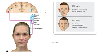
Bell palsy vs facial palsy due to stroke

Image
  Bell palsy vs facial palsy due to stroke   The two most common causes of acute facial paralysis are Bell’s palsy and ischemic stroke Facial weakness can be caused by strokes in many different locations in the brain and brainstem. Strokes involving the brain typically cause central facial weakness that involves the mouth and spares the eye and forehead. Strokes involving the brainstem can sometimes cause weakness of the mouth, eye and forehead–mimicking a peripheral lesion. In these cases however, there will be other focal neurologic deficits. A review of systems and neurologic examination can help to identify signs and symptoms of stroke. Bell's palsy is  a condition that causes sudden weakness in the muscles on one side of the face . In most cases, the weakness is temporary and significantly improves over weeks. The weakness makes half of the face appear to droop. Smiles are one-sided, and the eye on the affected side resists closing. Bell palsy vs facial palsy due to stroke  

difference between rough endoplasmic reticulum and smooth endoplasmic reticulum

Image
difference between rough endoplasmic reticulum and smooth endoplasmic reticulum

Difference between somitomere and somite of paraxial mesoderm

  Difference between somitomere and somite Somitomere Somite Loosely organized segmented collections of paraxial mesoderm in the cranial region Epithelial balls of cells formed in segmented pairs along the neural tube from paraxial mesoderm It form muscles and bones of the face and skull It forms vertebrae, muscles of the back and body wall and dermis of skin It has no subdivision It has three subdivision dermatome myotome sclerotome It is situated cranially It is situated caudally

Difference between general sensation and special sensation

  Difference between general sensation and special sensation General sensation Special sensation •       Touch •       Temperature •       Pain •       Pressure •       Proprioception from muscles & joint   •       Vibration   •       Itching ( is a subthreshold sensation of pain; both itch &  pain sensations share common nerve pathways) •       Visceral sensation •       Chemical •       Olfaction •       Taste •       Vision •       Balance •       Hearing   Functional component of general sensation General somatic afferent General visceral afferent Functional component of special   sensation Special   somatic afferent Special visceral afferent General somatic afferent : collect information from external world   General visceral afferent: collect information about internal organs of body   Special   somatic afferent : vision, b

Difference between alar plate and basal plate of the mantle layer of neural tube

  Difference between alar plate and basal plate  of the mantle layer of neural tube Alar plate Basal plate The  alar plates  (or  alar lamina ) is a neural structure in the embryonic nervous system. Sensory area in the dorsal region of the spinal cord and brain In the developing  nervous system , the  basal plate  is the region of the  neural tube  ventral to the  sulcus limitans they develops from dorsal aspect of mantle layer of neural tube they develops from ventral   aspect of mantle layer of neural tube Functional components deal by alar plate :    Cells within the alar plate develop into afferent and interneurons. The alar plate further develops general sensory afferents (GSA) innervating the skin, special sensory afferents (SSA) innervating the branchial arch derivatives, and visceral sensory afferents (VSA) innervating the respiratory, cardiac, and gastrointestinal systems.